Maxillary nerve (CN V2) Anatomy and function Kenhub
The maxillary nerve
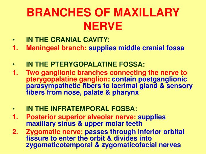
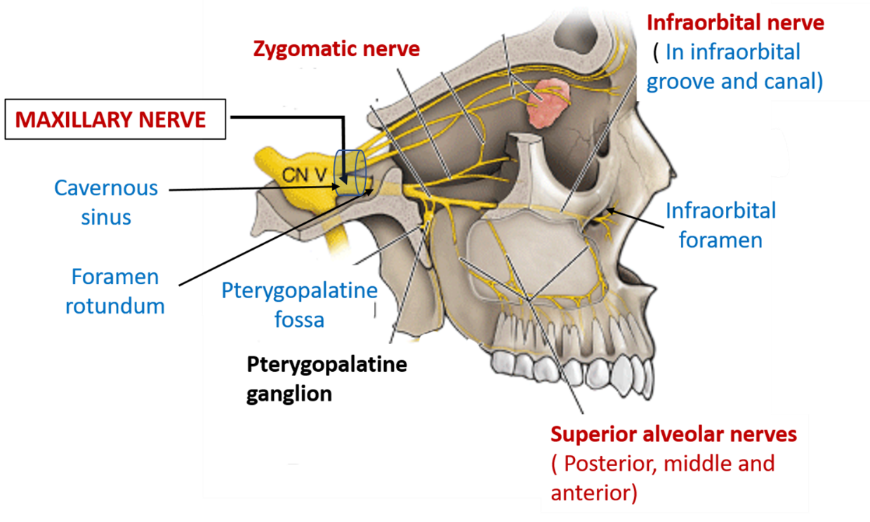
Maxillary nerve is the 2nd division of trigeminal nerve. It is a pure sensory nerve. It commences from the anterior aspect of trigeminal ganglion. It passes along the lateral wall of the cavernous sinus. It then leaves the cranial cavity via foramen rotundum and enter pterygopalatine fossa.

Maxillary nerve branches 4 YouTube
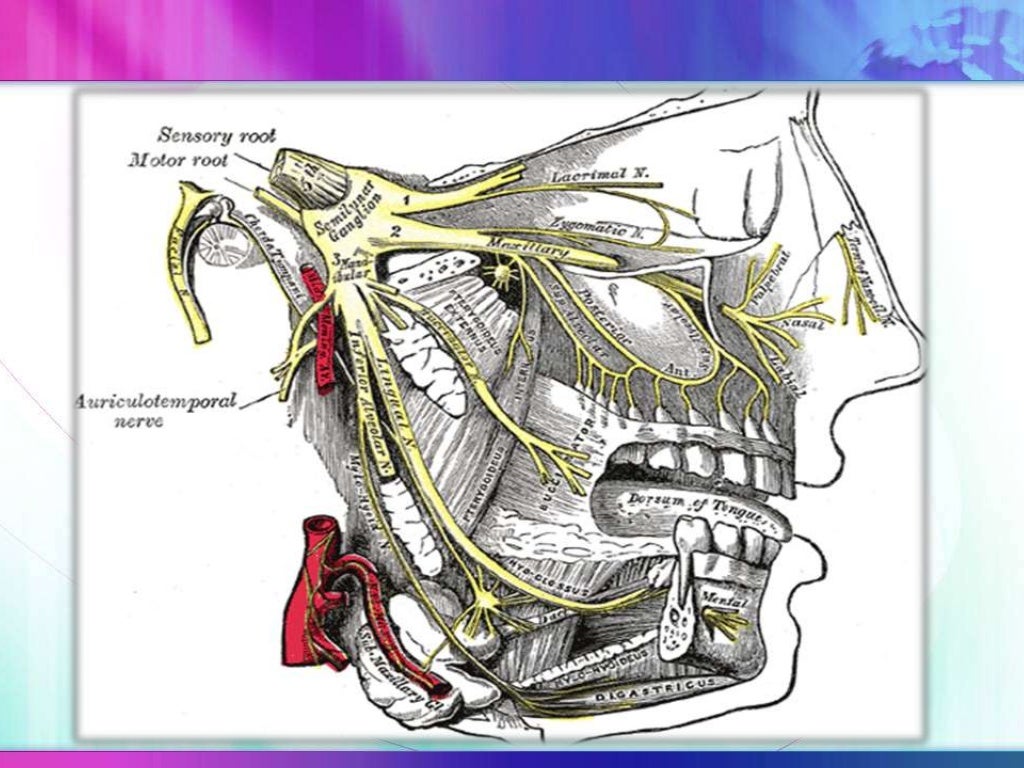
Maxillary Nerve. The maxillary nerve, or second division of the trigeminal, is a sensory nerve that crosses the pterygopalatine fossa, traverses the orbit in the infraorbital groove and canal in the floor of the orbit, and appears upon the face at the infraorbital foramen as the infraorbital nerve. From: Cosmetic Facial Surgery, 2011.
Superior Maxillary Nerve ClipArt ETC
As we've seen, the maxillary nerve runs forwards from the trigeminal ganglion, and enters the foramen rotundum, which is here. Here's the foramen rotundum in the dry bone. We'll go round to the outside to see where it emerges. Here it is, well hidden in the pterygo-maxillary fissure.

Anatomy and clinical significance of the maxillary nerve a literature review. Semantic Scholar
The maxillary nerve (V2) is the middle sized branch of the trigeminal nerve - the largest of the cranial nerves. The V2 is a purely sensory nerve supplying the maxillary teeth and gingiva, the.
Branches Of Maxillary Nerve slidesharedocs
The maxillary nerve is one of the branches of the trigeminal nerve, otherwise known as the fifth cranial nerve (CN V). Supplying sensory innervation to certain parts of the face, the mucosa of the nose, together with the teeth, this nerve allows you to feel that annoying fly landing underneath your eye or that annoying pain caused by your dentist.
PPT INFRATEMPORAL FOSSA II MAXILLARY NERVE & VESSELS PowerPoint Presentation ID1377415
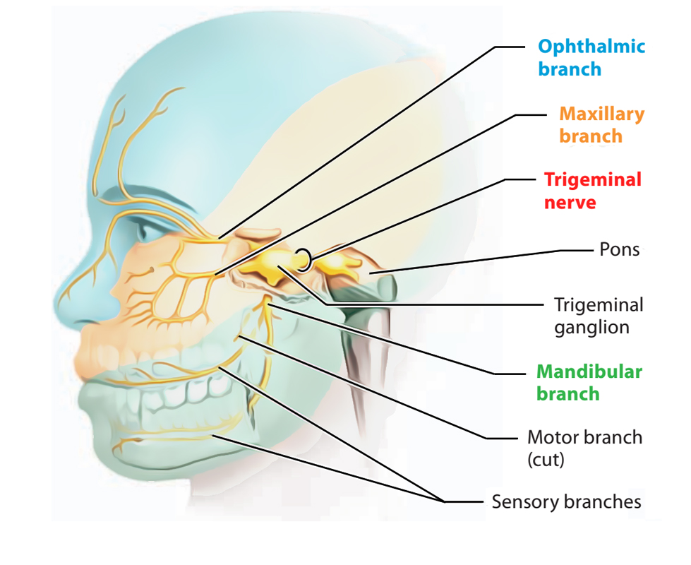
The maxillary nerve is the second of three branches of the trigeminal nerve. It arises between the trigeminal's ophthalmic and mandibular divisions in a region called the trigeminal ganglion, a cluster of nerves involved in relaying sensory information to the brain as well as chewing motor function.

[PDF] A review of the mandibular and maxillary nerve supplies and their clinical relevance
1/3 Synonyms: Antrum of Highmore, Maxillary paranasal sinus , show more. The maxillary sinus is the largest paranasal sinus situated in the body of the maxilla. The maxillary sinus is connected with the middle nasal meatus via the maxillary ostium. The maxillary sinus is bordered by three main walls:
Maxillary Nerve , Origin, Course and Branches , Anatomy QA
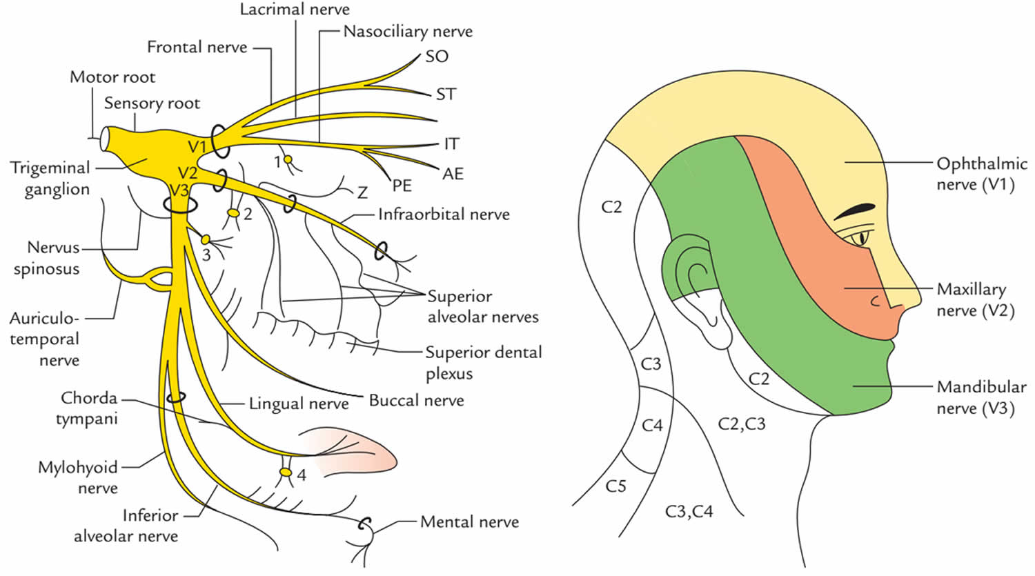
PMID: 31194417 Bookshelf ID: NBK542277 Excerpt The fifth cranial nerve, known as the trigeminal nerve (V), is the largest of the twelve cranial nerves and carries both sensory and motor fibers. It has three terminal branches, which in descending order are ophthalmic nerve (V1), maxillary nerve (V2), and mandibular nerve (V3).

Diagram of the second branch (maxillary) of the trigeminal nerve with its branches. Nerve
The maxillary sinus is intimately related to the roots of the posterior maxillary teeth; the high frequency of mucosal disease and sinusitis of odontogenic aetiology is now well recognized.
Maxillary nerve (CN V2) Anatomy and function Kenhub
The maxilla, also known as the upper jaw, is a vital viscerocranium structure of the skull. It is involved in the formation of the orbit, nose and palate, holds the upper teeth and plays an important role for mastication and communication.

Maxillary Nerve Lateral View Diagram Quizlet
Maxillary nerve is the 2nd branch of the trigeminal nerve. INTRODUC. Let's learn the course and branches of the maxillary nerve in this video in a unique way.

Maxillary nerve Branches, course (preview) Human Anatomy Kenhub YouTube
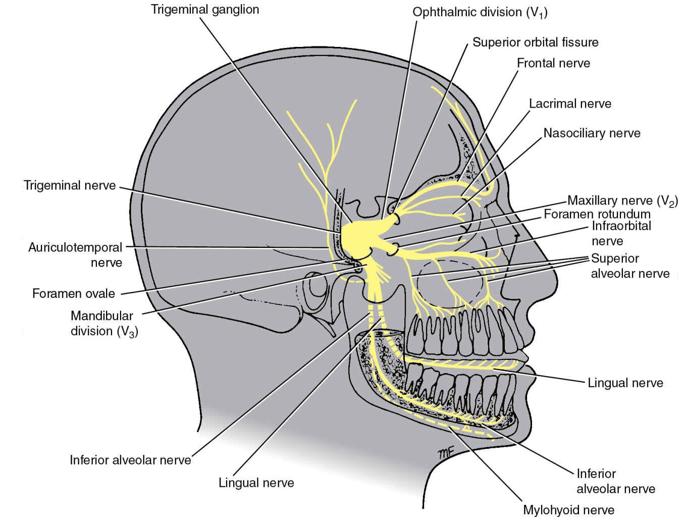
The maxillary nerve emerges from the anterior portion of the trigeminal ganglion, just inferior to the ophthalmic nerve. It runs anteriorly, passing through the lateral portion of the cavernous sinus until it reaches the foramen rotundum. It passes through the foramen rotundum to enter the upper pterygopalatine fossa.

Figure 1 from Anatomy and clinical significance of the maxillary nerve a literature review
The maxillary nerve arises from the anterior convexity of trigeminal ganglion between ophthalmic and mandibular divisions of the trigeminal nerve. It is a medium-sized branch compared to the smaller ophthalmic nerve and the larger mandibular nerve.
FIFTH CRANIAL NERVETRIGEMINAL NERVE
The maxillary nerve (CN V2, Latin: nervus maxillaris) is the second branch or division of the trigeminal nerve (CN V), also known as the maxillary division of the trigeminal nerve.

Maxillary nerve branches 2 YouTube
The Maxillary Nerve [Vb; V2] (n. maxillaris; superior maxillary nerve), or second division of the trigeminal, is a sensory nerve. It is intermediate, both in position and size, between the ophthalmic and mandibular. It begins at the middle of the semilunar ganglion as a flattened plexiform band, and, passing horizontally forward, it leaves the skull through the foramen rotundum, where it.
The Cranial Nerves Radiology Key
The maxillary nerve is the second branch of the trigeminal nerve, which originates embryologically from the first pharyngeal arch. Its primary function is sensory supply to the mid-third of the face. In this article, we shall look at the anatomy of the maxillary nerve - its anatomical course, sensory and parasympathetic functions. Anatomical Course
